Vegetos

(An International Journal of Plant Research & Biotechnology)
(eISSN: 2229-4473)
In silico analysis of Phlogacanthus thyrsiflorus Nees. and its protective effects against cardiomyopathic stress induced by alloxan in mice model
*Article not assigned to an issue yet
Research Articles | Published: 05 April, 2025
First Page: 0
Last Page: 0
Views: 1178
Keywords: Phlogacanthus thyrsiflorus, Methanolic flower extract, Alloxan, Hyperglycemia, Oxidative stress, Apoptosis
Abstract
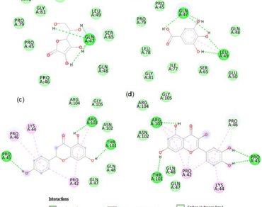
This study aims to evaluate the protective role of Phlogacanthus thyrsiflorus (P. thyrsiflorus) on hyperglycemia induced oxidative stress and apoptosis in the hearts of diabetic mice. Acute toxicity testing and preliminary phytochemical screens were performed on methanolic flower extract (MFE) after it had been prepared. Thereafter, molecular docking was performed. The results identified active compounds which demonstrated strong binding affinity with the selected target proteins involved in oxidative stress under diabetic conditions. Moreover, the oxidative damage in the tissues of the Normal control mice (NCM), Diabetic control mice (DCM), Ascorbic acid-treated Diabetic mice (D + AA), and MFE-treated Diabetic mice (D + MFE) groups were assessed using lipid peroxidation and the protein carbonyl test. The diabetic mice treated with MFE showed a significant reduction in protein carbonyl levels and malondialdehyde (MDA). The effect of MFE on apoptosis was demonstrated by TUNEL enzyme where a few apoptotic cells were shown by the terminal deoxynucleotidyl transferase (TdT)-mediated dUTP nick-end labeling (TUNEL) assay. Therefore, in diabetic mice, the MFE of P. thyrsiflorus has cardiomyopathy benefits via controlling hyperglycemia-induced oxidative stress and apoptosis.
References
Baird L, Yamamoto M (2020) The molecular mechanisms regulating the KEAP1-NRF2 pathway. Mol Cell Biol 40(13):e00099-e120. https://doi.org/10.1128/MCB.00099-20
Bora J, Sahariah P, Patar AK, Syiem D, Bhan S (2018) Attenuation of diabetic hepatopathy in alloxan-induced diabetic mice by methanolic flower extract of PhlogacanthusthyrsiflorusNees. J Appl Pharm 8(7):114–120
Bora J, Syiem D, Bhan S (2019) Quantitative analysis of total phenolic, flavonoid contents and HPTLC fingerprinting of flower extracts of Phlogacanthus thyrsiflorus nees. Journal of Pharmacognosy and Phytochemistry 8(3):906–911
Dalle-Donne I, Rossi R, Giustarini D, Milzani A, Colombo R (2003) Protein carbonyl groups as biomarkers of oxidative stress. Clin Chim Acta 329:23–38
Danilova IG, Sarapultsev PA, Medvedeva SU, Getti IF, Bulavintceva TS, Sarapultsev AP (2015) Morphological restructuring of myo cardium during the early phase of experimental diabetes mellitus. Anat Rec 298(2):396–407
Davies TG, Wixted WE, Coyle JE, Griffiths-Jones C, Hearn K, McMenamin R, Norton D, Rich SJ, Richardson C, Saxty G, Willems HM, Woolford AJ, Cottom JE, Kou JP, Yonchuk JG, Feldser HG, Sanchez Y, Foley JP, Bolognese BJ, Logan G, Kerns JK (2016) Monoacidic inhibitors of the Kelch-like ECH-associated protein 1: nuclear factor erythroid 2-related factor 2 (KEAP1:NRF2) protein-protein interaction with high cell potency identified by fragment-based discovery. J Med Chem 59(8):3991–4006. https://doi.org/10.1021/acs.jmedchem.6b00228
Draper HH, Hadley M (1990) Malondialdehyde determination as index of lipid peroxidation. Meth Enzymol 186:421–431
El-Far AH, Sroga G, Al Jaouni SK, Mousa SA (2020) Role and mechanisms of RAGE-ligand complexes and RAGE-inhibitors in cancer progression. Int J Mol Sci 21(10):3613
Forli S, Huey R, Pique ME, Sanner MF, Goodsell DS, Olson AJ (2016) Computational protein-ligand docking and virtual drug screening with the AutoDock suite. Nat Protoc 11(5):905–919. https://doi.org/10.1038/nprot.2016.051
Galicia-Garcia U, Benito-Vicente A, Jebari S, Larrea-Sebal A, Siddiqi H, Uribe KB, Ostolaza H, Martín C (2020) Pathophysiology of Type 2 diabetes mellitus. Int J Mol Sci 21(17):6275. https://doi.org/10.3390/ijms21176275
Graham J (2002) Preparation of crude subcellular fractions by differential centrifugation. Sci WorldJ 2:1638–1642
Jena AB, Samal RR, Bhol NK, Duttaroy AK (2023) Cellular Red-Ox system in health and disease: The latest update. Biomed Pharmacother 162:114606
Kay AM, Simpson CL, Stewart JA (2016) The role of AGE/RAGE signaling in diabetes-mediated vascular calcification. J Diabetes Res 2016:1
Kim S, Thiessen PA, Bolton EE, Chen J, Fu G, Gindulyte A, Han L, He J, He S, Shoemaker BA, Wang J, Yu B, Zhang J, Bryant SH (2016) PubChem substance and compound databases. Nucleic Acids Res 44(D1):D1202–D1213. https://doi.org/10.1093/nar/gkv951
Kumar SS, Devasagayam TPA, Bhushan B, Verma NC (2001) Scav enging of reactive oxygen species by chlorophyllin: an ESR study. Free Radic Res 35:563–574
Lee KS, Buck M, Houglum K, Chojkier M (1995) Activation of hepatic stellate cells by TGF alpha and collagen type I is medi ated by oxidative stress through c-myb expression. J Clin Invest 96:2461–2468
Lenzen S (2008) The mechanisms of alloxan- and streptozotocin induced diabetes. Diabetologia 51(2):216–226
O’Boyle NM, Banck M, James CA, Morley C, Vandermeersch T, Hutchison GR (2011) Open babel: an open chemical toolbox. J Cheminform 3:33. https://doi.org/10.1186/1758-2946-3-33
Pan HZ, Zhang L, Guo MY et al (2010) The oxidative stress status in diabetes mellitus and diabetic nephropathy. Acta Diabetol 47:71–76
Parola M, Leonarduzzi G, Biasi F, Albano E, Biocca ME, Poli G, Dianzani MU (1992) Vitamin E dietary supplementation protects against carbon tetrachlorideinduced chronic liver damage and cir rhosis. Hepatology 16:1014–1021
Pizzino G, Irrera N, Cucinotta M, Pallio G, Mannino F, Arcoraci V, Squadrito F, Altavilla D, Bitto A (2017) Oxidative stress: harms and benefits for human health. Oxid Med Cell Longev 2017:8416763. https://doi.org/10.1155/2017/8416763
Pradeepa R, Mohan V (2021) Epidemiology of type 2 diabetes in India. Indian J Ophthalmol 69(11):2932–2938. https://doi.org/10.4103/ijo.IJO_1627_21
Author Information
Amity Institute of Biotechnology, Amity University Jharkhand, Ranchi, India