Vegetos

(An International Journal of Plant Research & Biotechnology)
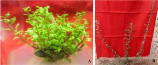
Repairing mechanism of foliar micro-morphological anomalies during acclimatization and field transfer of in vitro raised plantlets of Aerva lanata (L.) Juss. ex Schult.: a medicinally important plant
Research Articles | Published: 16 November, 2021
Online ISSN : 2229-4473.
Website:www.vegetosindia.org
Pub Email: contact@vegetosindia.org
First Page: 520
Last Page: 526
Views: 1723
Keywords:
Adaptation,
Aerva lanata
, Leaf micro-morphology, Micropropagation, Stomata
Abstract
The present report elucidates the foliar micro-morphological adaptation mechanism of micropropagated plantlets of Aerva lanata. The in vitro induced anomalies in the leaves were repaired through gradual hardening and acclimatization processes in a greenhouse. The epidermal tissues of in vitro grown leaves significantly differed from the field transferred plants in terms of architecture and undulations of anticlinal walls. Amphistomatic leaves with anomocytic stomata were observed with the in vitro cultured and field transferred plants, but the stomata were significantly different in their morphology, size, and spatial distribution. The in vitro leaves possessed high stomatal density (59.4) and stomatal index (28.9), but a significant decrease in stomatal density (48.7) and stomatal index (26.3) were observed in the field established plants. Vein density, vein-islets, and vein-let terminations of the leaves were increased from in vitro to field environments. The trichomes of in vitro leaves were multicellular, uniseriate, and underdeveloped which attained gradual maturity when transferred to the field. The calcium oxalate rosette crystals were unorganized, underdeveloped, and varied in size with the in vitro leaves; these were organized, completely developed, and morphologically uniform in acclimatized leaves. The study revealed that the gradual changes in foliar micro-morphological parameters such as stomatal apparatus, trichomes, crystals, and vein density from in vitro to in vivo conditions involved in the repair mechanism of the photosynthetic organs could increase the survival chances of micropropagated plantlets in the field.
(*Only SPR Members can get full access. Click Here to Apply and get access)
References
Afreen F (2005) Physiological and anatomical characteristics of in vitro photoautotrophic plants. In: Kozai T, Afreen F, Zobayed S (eds) Photoautotrophic (sugar-free medium) micropropagation as a new micropropagation and transplant production system. Springer, Dordrecht, pp 61–90. https://doi.org/10.1007/1-4020-3126-2_6
Bairu MW, Kane ME (2011) Physiological and developmental problems encountered by in vitro cultured plants. Plant Growth Regul 63:101–103
Bairu MW, Aremu AO, Van Staden J (2011) Somaclonal variation in plants: causes and detection methods. Plant Growth Regul 63:147–173
Berger D, Altmann T (2000) A subtilisin-like protease involved in the regulation of stomatal density and distribution in Arabidopsis thaliana. Genes Dev 14:1119–1131
Camargo MAB, Marenco RA (2011) Density, size and distribution of stomata in 35 rainforest tree species in Central Amazonia. Acta Amaz 41:205–212
Croxdale JL (2000) Stomatal patterning in angiosperms. Am J Bot 87:1069–1080
George EF (2008) Plant tissue culture procedure—background. In: George EF, Hall MA, De KlerK GJ (eds) Plant propagation by tissue culture. Springer, Dordrecht, pp 1–28
Hanley ME, Lamont BB, Fairbanks MM, Rafferty CM (2007) Plant structural traits and their role in anti-herbivore defence. Perspect Plant Ecol Evol Syst 8:157–178
Hickey LJ, Wolfe JA (1975) The bases of angiosperm phylogeny. Vegetative morphology. Ann Missouri Bot Gard 62:538–589
Iliev I, Kitin P (2011) Origin, morphology and anatomy of fasciation in plants cultured in vivo and in vitro. Plant Growth Regul 63:115–129
Isah T (2015) Adjustments to in vitro culture conditions and associated anomalies in plants. Acta Biol Cracoviensia Ser Bot 57:9–28
Jogam P, Sandhya D, Shekhawat MS, Alok A, Manokari M, Abbagani S, Allini VR (2020) Genetic stability analysis using DNA barcoding and molecular markers and foliar micro-morphological analysis of in vitro regenerated and in vivo grown plants of Artemisia vulgaris L. Ind Crops Prod. https://doi.org/10.1016/j.indcrop.2020.112476
Johansen DA (1940) Plant microtechnique. McGraw Hill Book Company, New York, pp 182–197
Kher MM, Nataraj M (2017) Micropropagation of Combretum ovalifolium Roxb.: a medicinally important plant. Rend Fis Acc Lincei Doi. https://doi.org/10.1007/s12210-017-0625-z
Kostman TA, Tarlyn NM, Loewus FA, Franceschi VR (2001) Biosynthesis of L-ascorbic acid and conversion of carbons 1 and 2 of L-ascorbic acid to oxalic acid occurs within individual calcium oxalate crystal idioblasts. Plant Physiol 125:634–640
Kozai T, Xiao Y (2006) A commercialized photoautotrophic micropropagation system. In: Gupta SD, Ibaraki Y (eds) Plant tissue culture engineering. Springer, Dordrecht, pp 355–371
Machado MP, Silva ALL, Biasi LA, Deschamps C, Filho JCB, Zanette F (2014) Influence of calcium content of tissue on hyperhydricity and shoot tip necrosis of in vitro regenerated shoots of Lavandula angustifolia Mill. Braz Arch Biol Technol 57:636–643
Magyar-Tábori K, Dobránszki J, Teixeira da Silva JA, Bulley SM, Hudák I (2010) The role of cytokinins in shoot organogenesis in apple. Plant Cell Tiss Org Cult 101:251–267
Manokari M, Shekhawat MS (2017) Optimization of in vitro and ex vitro regeneration and micromorphological studies of Micrococca mercurialis (L.) Benth. Bot Pac 6(1):37–44
Moyo M, Aremu AO, Van Staden J (2015) Insights into the multifaceted application of microscopic techniques in plant tissue culture systems. Planta 242:773–790
Murashige T, Skoog F (1962) A revised medium for rapid growth and bioassays with Tobacco tissue culture. Physiol Plant 15:473–497
Nagaratna A, Hegde PL, Harini A (2015) A Pharmacological review on Gorakha ganja (Aerva lanata (Linn) Juss. Ex. Schult). J Pharmacogn Phytochem 3:35–39
Nandagopal S, Lalitha M, Abirami S, Saikrishna D, Priyan A (2015) Effect of phytohormones on micropropagation and phytochemical studies of Aerva lanata (Linn.) Juss.ex Schult—a seasonal and vulnerable plant. Der Pharm Lett 7:291–298
Pence VC (2010) The possibilities and challenges of in vitro methods for plant conservation. Kew Bull 65:539–547
Pospisilova J, Ticha I, Kadlecek P, Haisel D, Plzakova S (1999) Acclimatization of micropropagated plants to ex vitro conditions. Biol Plant 42:481–497
Prabhakar M (2004) Structure, delimitation, nomenclature and classification of stomata. Acta Bot Sin 46:242–252
Ramachandra YL, Shilali K, Mansoor A, Sudeep HV, Kavitha BT, Gurumurthy H, Rai SP (2011) Hepatoprotective properties of Boerhaavia Diffusa and Aerva lanata against carbon tetra chloride induced hepatic damage in rats. Pharmacologyonline 3:435–441
Revathi J, Manokari M, Latha R, Priyadharshini S, Kher MF, Shekhawat MS (2019) In vitro propagation, in vitro flowering, ex vitro root regeneration and foliar micro-morphological analysis of Hedyotis biflora (Linn.) Lam. Vegetos 32:609–619
Revathi J, Manokari M, Priyadharshini S, Shekhawat MS (2020) Foliar micromorphometric adaptations of micropropagated plants of Oldenlandia herbacea (L.) Roxb.—an important medicinal herb. Curr Sci 119(2):398–401. https://doi.org/10.18520/cs/v119/i2/398-401
Sahay NS, Varma A (2000) A biological approach towards increasing the rates of survival of micropropagated plants. Cur Sci 78:126–129
Sahu AR, Rath SC, Panigrahi J (2013) In vitro propagation of Aerva lanata (l.) Juss. ex Schult. through organogenesis. Indian J Biotechnol 12:260–264
Salisbury EJ (1932) The interrelations of soil climate and organisms and the use of stomatal frequency as an integrating index of relation of the plant. Bech Bot Zbl 99:402–420
Sandhya D, Jogam P, Manokari M, Shekhawat MS, Jadaun JS, Allini VR, Abbagani S (2021) High frequency in vitro propagation and assessment of genetic uniformity and micro-morphological characterization of Origanum majorana L.—a highly traded aromatic herb. Biocat Agric Biotechnol. https://doi.org/10.1016/j.bcab.2021.102024
Shekhawat MS, Manokari M (2016a) In vitro propagation, micromorphological studies and ex vitro rooting of cannon ball tree (Couroupita guianensis aubl.): a multipurpose threatened species. Physiol Mol Biol Plants 22:131–142
Shekhawat MS, Manokari M (2016) Optimization of in vitro and ex vitro regeneration and micromorphological studies in Basella alba L. Physiol Mol Biol Plants 22:605–612
Shekhawat MS, Manokari M (2018) Micromorpho-anatomical evaluation of in vitro and field transferred plants of Coccinia indica Wight and Arn. Agric Res 7:135–144. https://doi.org/10.1007/s40003-018-0326-6
Shekhawat MS, Manokari M, Revathi J (2016) In vitro propagation and ex vitro rooting of Aerva lanata (L.) Juss. ex Schult.: a rare medicinal plant. Ind J Plant Physiol. https://doi.org/10.1007/s40502-016-0248-x
Stefanova MA, Koleva DP (2014) Leaf’s structural response of in vitro cultured Leonurus cardiaca plants to N6-benzyladenine and indole-3-butyric acid. J Pharm Res B 8:1014–1021
Sudeesh S (2012) Ethnomedicinal plants used by Malayaraya tribes of Vannapuram village in Iduki, Kerala, India. Indian J Sci Res Technol 1:7–11
Valverde PL, Fornoni J, Nunez-Farfan J (2001) Defensive role of leaf trichomes in resistance to herbivorous insects in Datura stramonium. J Evol Biol 14:424–432
van Staden J, Fennell CW, Taylor NJ (2006) Plant stress in vitro: the role of phytohormones. In: Fári MG, Holb I, Bisztray GD (eds) Proceedings of Vth IS on in vitro culture and horticulture breeding, ISHS Acta Hort vol 725, pp 55-61. https://doi.org/10.17660/ActaHortic.2006.725.2
Varutharaju K, Raju CS, Thilip C, Aslam A, Shajahan A (2014) High efficiency direct shoot organogenesis from leaf segments of Aerva lanata (L.) Juss. ex Schult by using Thidiazuron. Sci World J. https://doi.org/10.1155/2014/652919
Zhao Y, Kumar D, Prasad DN, Singh RK, Ma Y (2015) Morphoanatomic, physicochemical, and phytochemical standardization with HPTLC fingerprinting of aerial parts of Aerva lanata (Linn) Juss ex Schult. J Tradit Chin Med Sci. https://doi.org/10.1016/j.jtcms.2014.12.002
Acknowledgements
Authors are grateful to the Science and Engineering Research Board, Department of Science and Technology and National Medicinal Plants Board, Ministry of AYUSH, Government of India for providing financial support to their laboratory under the EMR Scheme Grant no. EMR/2016/007795 and NMPB/IFD/GIA/NR/PL/2018-19/187 respectively.
Author Information
Department of Botany, Siddha Clinical Research Unit, Central Council for Research in Siddha, Palayamkottai, India
Biotechnology Unit, Kanchi Mamunivar Government Institute for Postgraduate Studies and Research, Puducherry, India
Biotechnology Unit, Kanchi Mamunivar Government Institute for Postgraduate Studies and Research, Puducherry, India
smahipal3@gmail.com